Diagnostic/ Nuclear Medicine
Nuclear Medicine
Diagnostic/ Nuclear Medicine
Nuclear Medicine
Tumour imaging is an integral part of cancer management. Our expert team of Nuclear Medicine Physicians and Technologists have the vast experience in that. Here, we give the commitment of delivering the quality images and interpretation, for the best outcome of your illness.
Get a call back from our health advisor
How Does PET-CT Work?

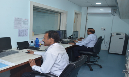
A special camera is used to detect these radioactive emissions and produce images of the metabolic activity of the scanned region. A CT scan that is carried out simultaneously captures the X-ray images of internal organs from different angles.
A computer then combines the data from PET and CT scans to produce 3-dimensional images of internal organs. Any abnormalities, like tumors, can be easily and accurately detected through a PET-CT scan.
Advantages of PET-CT Scan
PET-CT imaging has numerous advantages:
- Through enhanced accuracy, PET-CT supports the right diagnosis and, thereby, the right clinical decisions.
- PET-CT helps specialists detect cancers in their early stages, when they can be treated with positive outcomes.
- By providing highly reliable information about the lesions or abnormal masses, PET-CT imaging helps in effective treatment planning.
- PET-CT imaging also helps assess the patient’s response to the treatment given.
- In some cases, PET-CT can help prevent unnecessary invasive procedures and retests by providing helpful, comprehensive molecular and structural data at once.
- This imaging process is non-invasive, and in most cases, there will be no downtime or recovery required for the patients after the test.
- PET-CT imaging also plays an important role in radiotherapy planning.
Schedule your appointment


